
Brain inferior view, Part of the central nervous system enclosed in
Original upload log [edit]. File:Brain_human_normal_inferior_view.svg licensed with Cc-by-2.5 . 2009-10-13T16:18:05Z Beao 424x505 (209117 Bytes) Replaced right brain half with a clone of left brain half because they look excly the same in the picture.; 2007-09-23T15:14:17Z Ysangkok 424x505 (417241 Bytes) removing credits; 2007-03-03T17:30:01Z Ysangkok 424x505 (417718 Bytes) trying to make it.
InferiorBrainModel
Learn about the features, markings, and distinguishing characteristics of the brain; then test yourself with labeled images, hints, and answer keys that put you in control. Structure-Function.org. Resources biology human anatomy ☰ Brain Model, inferior view « Inferolateral view | Brain main.
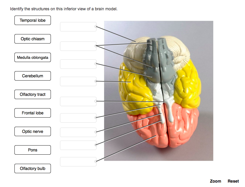
Solved Identify the structures on this inferior view of a
RM K2290P - Part of the central nervous system enclosed in the skull, consisting of the cerebrum, cerebellum and brain stem; it is responsible for sensory perception, most movements, memory, language, reflexes and vital functions. RF 2FYMW7M - Brain with highlighted inferior temporal gyrus, illustration
Brain Anatomy, Inferior View Photograph by Gwen Shockey Fine Art America
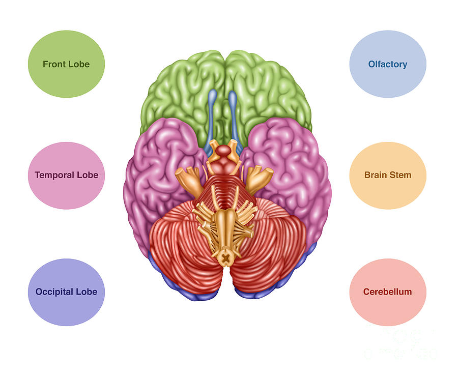
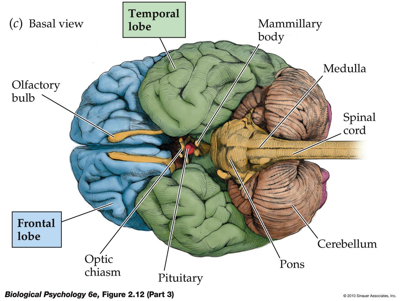
1/7 Synonyms: Forebrain, Endbrain , show more. The brain, along with the spinal cord, is the main organ of the central nervous system. It is the most complex organ of the body, with many layers and components that play their roles in almost every function performed by the body. The brain is composed of the cerebrum, cerebellum and brainstem.

Inferior brain Neuro, Neurology, Brain
Posterior Communicating Artery. Location. Term. Left Internal Carotid Artery. Location. Start studying Inferior View of the Brain. Learn vocabulary, terms, and more with flashcards, games, and other study tools.

Brain 🧠 inferior view Medical school studying, Nursing school
Genu of the corpus callosum (inferior view) The genu (Latin for knee) of the corpus callosum is observed in the center of the section, medial to the frontal lobes and the frontal (anterior) horns of the lateral ventricles .
Anatomy Of Human Brain, Inferior View Photograph by Alan Gesek Fine
Figure 1: Anatomy of the cranial base, inferior view. Figure 2: Anatomy of the hard palate and bony nasal septum, A. inferior view, and B. parasagittal view. Figure 3: Anatomy of the sphenoid bone, A. superior, B. inferior, and C. anterior views. Figure 4: Interior of the cranial base, superior view.
Cerebrum Overview
Inferior view Frontal lobe Temporal lobe Highlights Lateral view Medial view Inferior view Sources + Show all

Brain Anatomy, Inferior View, Illustration Stock Image F031/8230
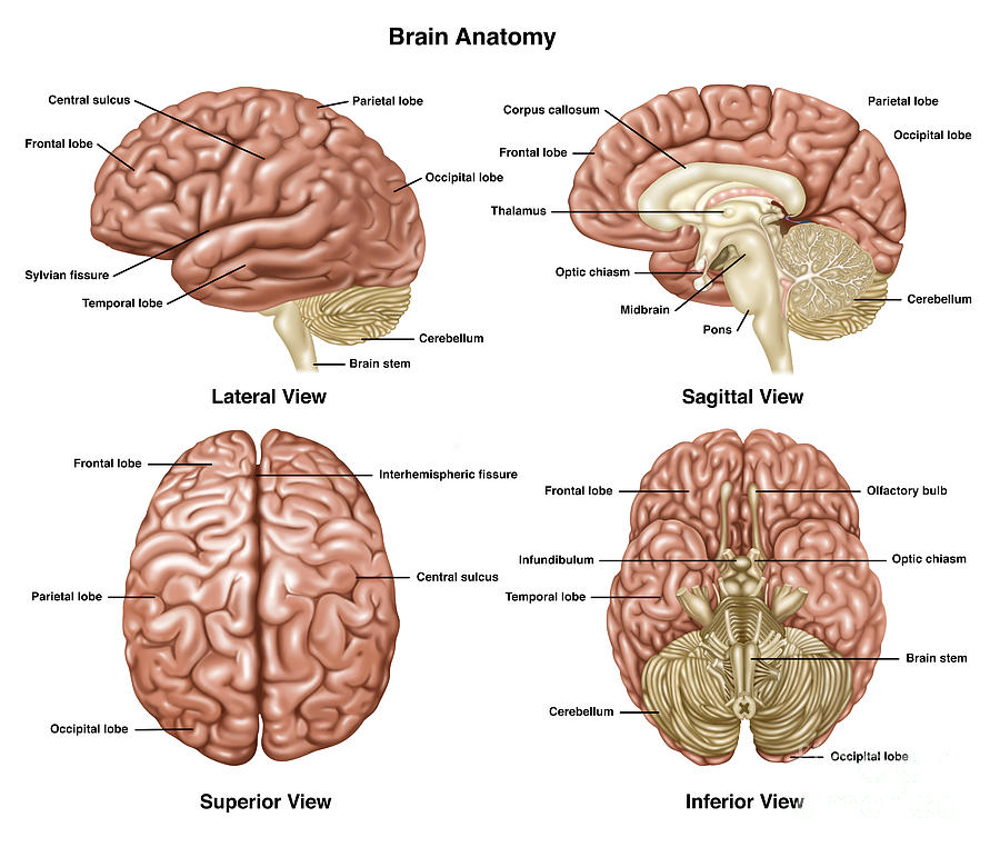
The lateral view of the brain shows the three major parts of the brain: cerebrum, cerebellum and brainstem . A lateral view of the cerebrum is the best perspective to appreciate the lobes of the hemispheres. Each hemisphere is conventionally divided into six lobes, but only four of them are visible from this lateral perspective.

The Brain Stem Basicmedical Key
Views of the brain: 1 2 3 4 5 6 Directions, Reference Planes, & Views of the Brain; explained beautifully in an illustrated and interactive way. Click and start learning now!
Brain Anatomy, Inferior View Photograph by Gwen Shockey
Identify the bones and structures that form the nasal septum and nasal conchae, and locate the hyoid bone. Identify the bony openings of the skull. The cranium (skull) is the skeletal structure of the head that supports the face and protects the brain. It is subdivided into the facial bones and the brain case, or cranial vault ( Figure 7.3 ).

Brain inferior view Brain models, Brain, Human brain
Cross sectional anatomy: MRI of the brain. An MRI was performed on a healthy subject, with several acquisitions with different weightings: spin-echo T1, T2 and FLAIR, T2 gradient-echo, diffusion, and T1 after gadolinium injection. We obtained 24 axial slices of the normal brain. Data and DICOM images archived on our PACS (Picture Archiving and.
Life After Being A Student My Mission To Learn The Brain
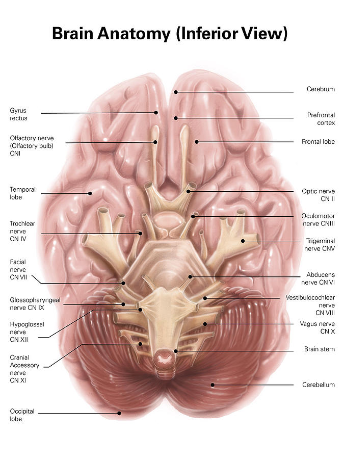
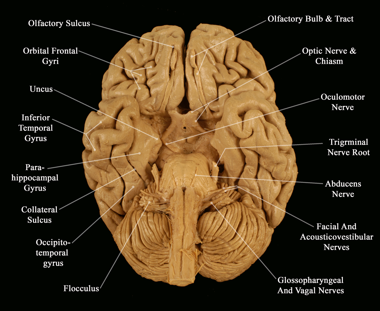
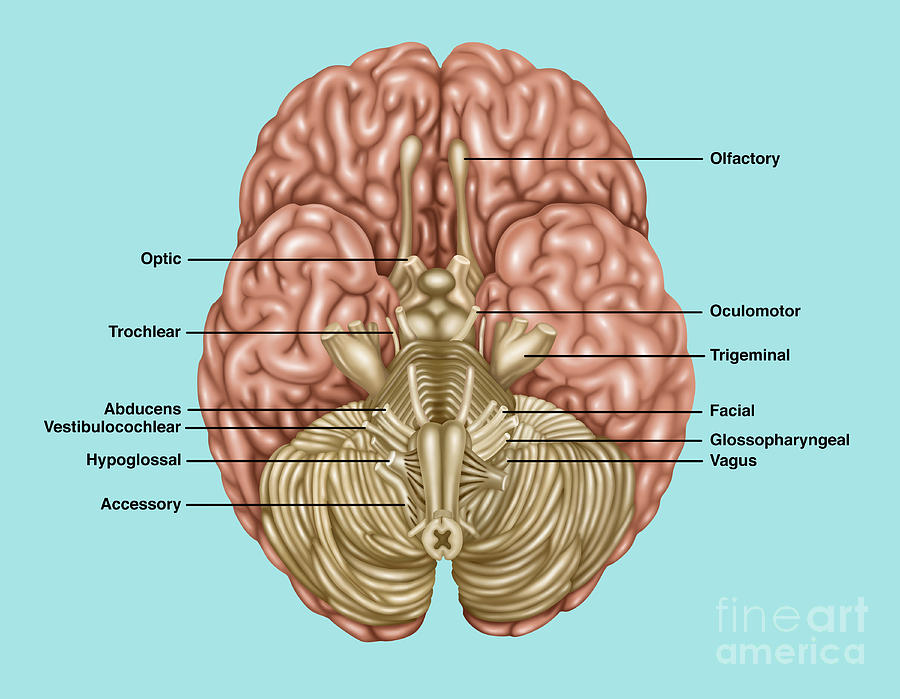
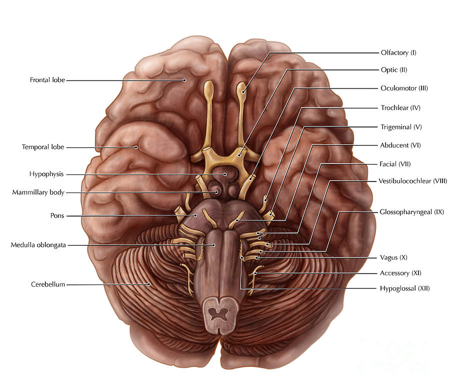
$300.00 Cranial nerves of the brain from an inferior (bottom) view. Add to cart Categories Anatomy, Head, Brain, Nervous Description This panel depicts the cranial nerves of the brain from an inferior (bottom) view.

7 Inferior view of arteries of the brain. Circle of Willis is depicted
The Brain - Inferior View. Create healthcare diagrams like this example called The Brain - Inferior View in minutes with SmartDraw. SmartDraw includes 1000s of professional healthcare and anatomy chart templates that you can modify and make your own. 62/75 EXAMPLES.
Brain Anatomy, Illustration Photograph by Gwen Shockey Fine Art America
Inferior View of the Base of the Cerebrum Tags Amygdala Anterior Cerebral Artery Anterior Choroidal Artery Basal Vein of Rosenthal Basilar Artery Cerebral Aqueduct Choroid Plexus Fimbria of Fornix Fusiform Gyrus Great Cerebral Vein of Galen Gyrus Rectus Infundibulum Internal Carotid Artery Interpeduncular Artery Lateral Posterior Choroidal Artery
Brain And Cranial Nerves Photograph by Evan Oto Fine Art America
Ways to View the Brain: the "nervous system" (including the brain) has several orientational directions.(Fisch, 4) It is common to combine terms. For example, a structure may be described as 'dorso-lateral,' which means that it is located 'up and to the side.' (Kolb, 39) A variety of terms is used for different directions and planes of section in the nervous system.
